
3D Simulation and Surgical Planning
At Prof. Dr. İlker Yazıcı's private practice in Nişantaşı, Istanbul, a surgical plan is not built on conversation and flat photographs alone. The face or torso is captured in 3D in the consultation room, the planned change is shown on screen with simulation software, and where useful the plan is 3D printed as a physical model and as a template used in the operating theatre. Patient and surgeon look at the same goal. This page explains how it works and where its limits lie.
How the 3D consultation works
For the relevant procedures, 3D imaging is part of the consultation, not a separate appointment. The camera system, the simulation software and the 3D printer serve one plan: the goal agreed on screen reaches the operating theatre through the printed model and template, where the shape achieved can be checked against it.
Phrases such as "a little more lifted" or "slightly fuller" mean different things to different people; on a shared screen they become a concrete shape. The simulation is also where unrealistic wishes are named: not every change drawn on screen can be achieved safely with your tissues. A consultation usually runs like this:
- Your goals: what you would like to change or keep, your medical history and any previous surgery.
- Examination: Prof. Dr. İlker Yazıcı examines the nose, chin or chest and assesses skin thickness, tissue elasticity, bone and cartilage support and asymmetry.
- 3D capture: the face or torso is photographed from several angles with the 3D surface-imaging camera system; it is quick and uses no X-rays. The images become a 3D model that can be rotated on screen.
- Simulation: the planned change is shown on your own 3D model, options are compared side by side and the goal is agreed.
- Plan: if surgery is appropriate, the written plan is discussed; for nose and breast surgery, 3D printed models and templates are prepared beforehand where useful.
Rhinoplasty: simulation, 3D printed model and surgical template
In rhinoplasty, 3D simulation shows the effect of reducing a dorsal hump, changing tip projection and rotation, adjusting the angle between nose and upper lip, and the overall balance of the profile, from the front, the side and below. Because the model rotates, the nose is judged against the whole face, not a single photograph, which is particularly useful for asymmetric or previously operated noses.
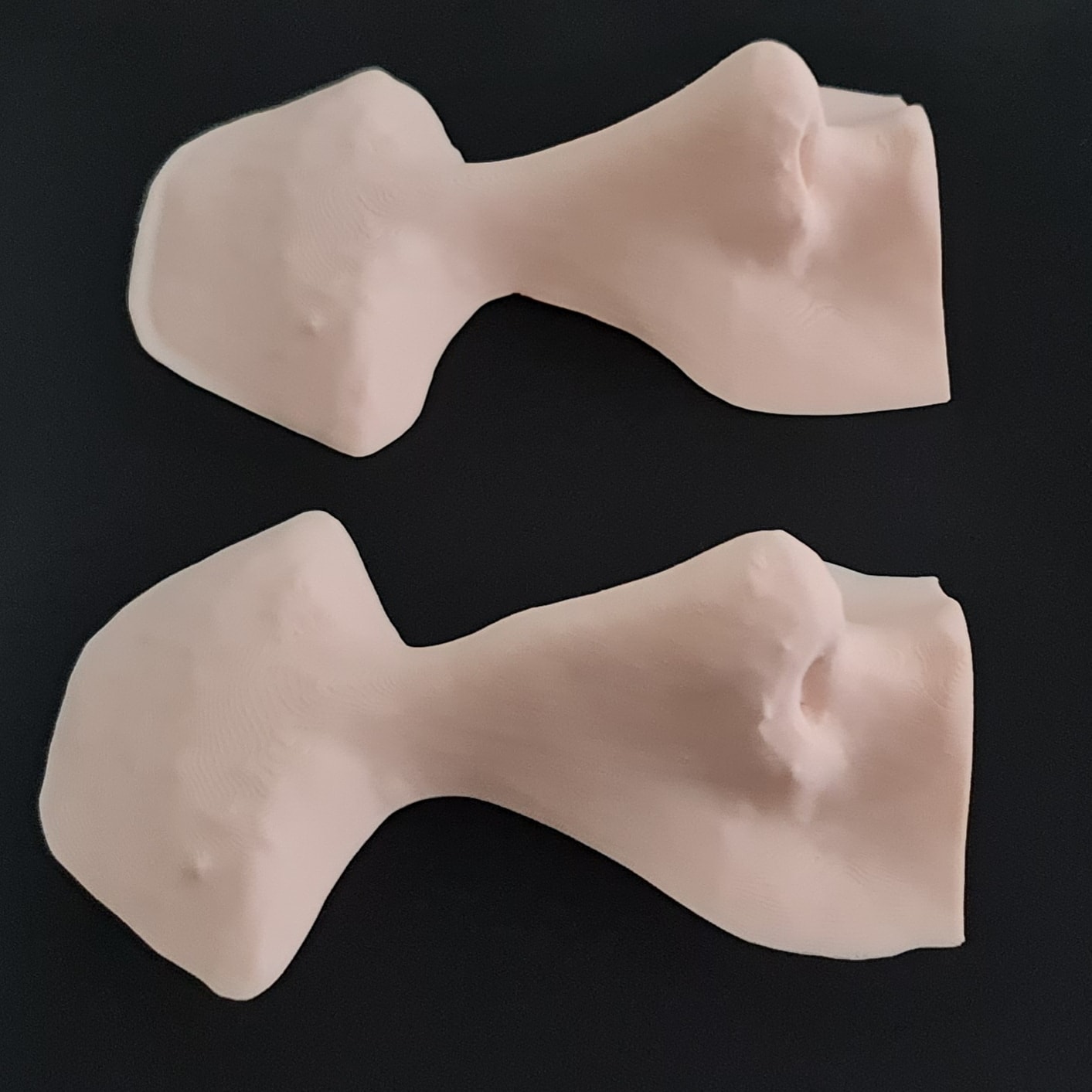
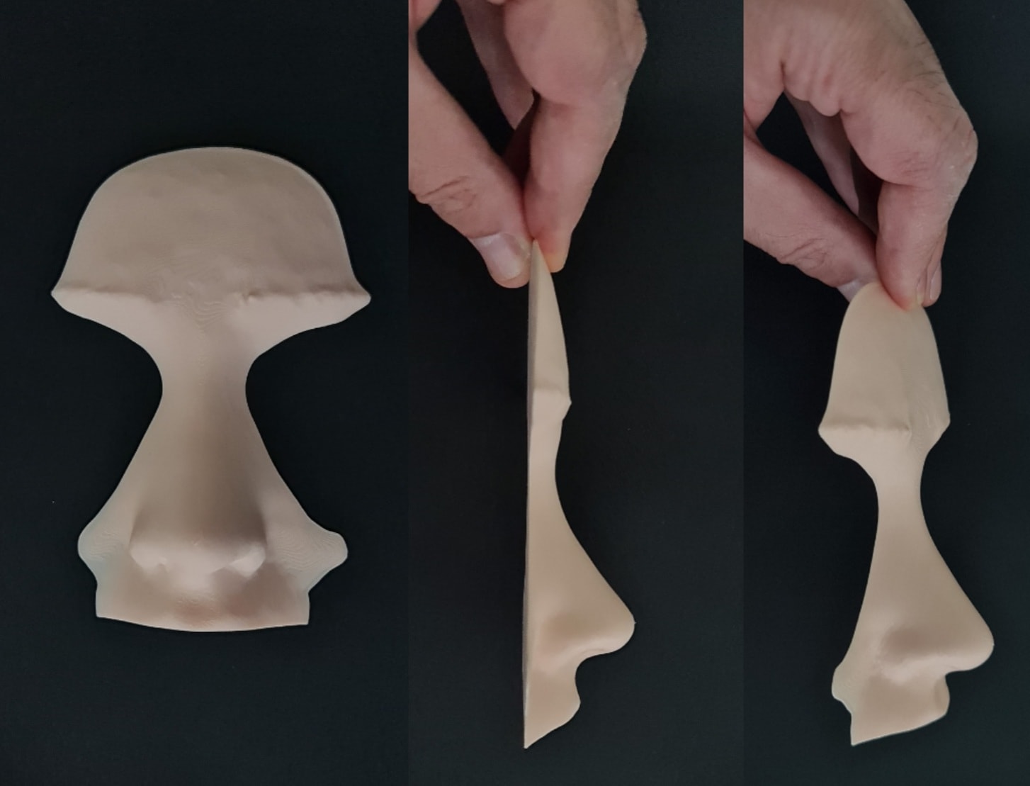
Where useful, the agreed simulation is 3D printed as a physical model of the planned nose, which you can hold and view from every angle. From the same plan, transparent patient-specific templates are printed. After sterilisation, the template is placed on the nose during surgery and the shape achieved at that moment is compared with the agreed plan.
The template is a checking tool; it does not replace surgical judgement. Swelling during the operation, cartilage strength and skin thickness are also weighed. In thicker skin, changes to the cartilage show less clearly on the surface, and this is discussed at the start of 3D rhinoplasty planning.
Breast surgery: volume and shape planning with simulation and 3D printed models
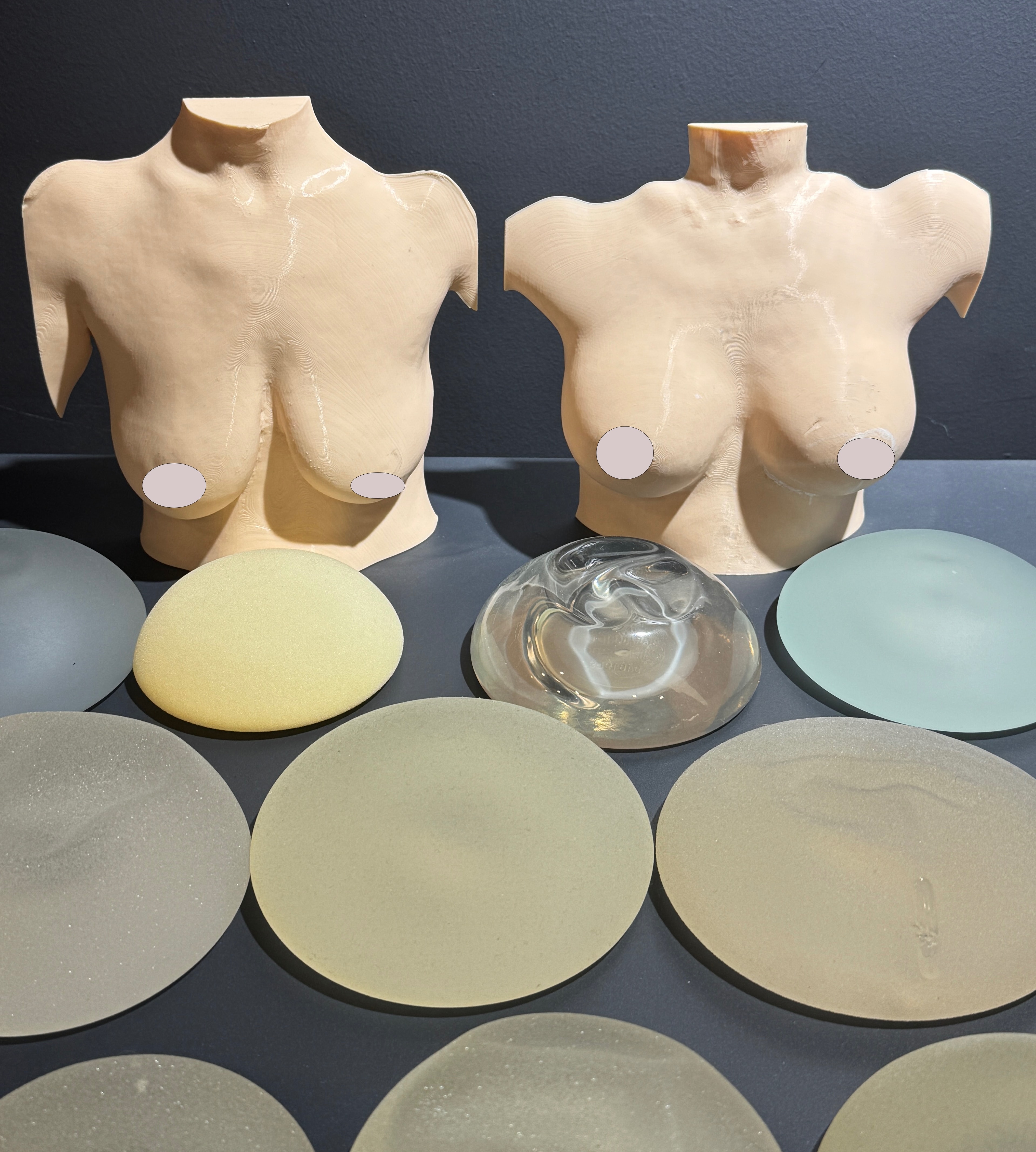
Before breast augmentation or a breast lift, the torso is captured in 3D. In 3D breast augmentation simulation, silicone implants of different volumes are tried on the model, and fullness, upper-pole shape, the gap between the breasts and the fit with the chest wall are compared from the front and side. When a lift is planned, the shape created by raising the nipple and breast tissue is discussed too.
The 3D model also shows differences in volume and position between the two breasts. Some asymmetry is normal, and a small difference pointed out before surgery is far less of a surprise afterwards.
3D printed torso models are used to talk about volume and shape; seeing a change on a physical model, not only on screen, makes the conversation about implant size more concrete. The final choice also depends on chest width, breast tissue, skin quality and lifestyle, together with the examination findings.
Chin and facial profile
The nose, lips and chin are seen together in profile. A recessed chin can make the nose look larger, so for some people considering rhinoplasty a chin correction (genioplasty) is assessed as well. The simulation can show how bringing the chin forward or reshaping it affects the profile, with or without the nose change.
The chin can be advanced by cutting and moving the chin bone (sliding genioplasty) or with a chin implant. The choice depends on bone structure, the bite and the amount of change needed, and is made at the examination. With a significant bite problem, chin surgery alone may not be enough and a different assessment is needed.
Reconstructive and maxillofacial planning from CT
For operations on bone, a surface image is not enough. In maxillofacial surgery and reconstructive surgery, computed tomography (CT) data is turned into 3D models of the skull and jaw, which can be studied on screen or 3D printed and handled before the operation.
For an orbital floor fracture (the floor of the eye socket), for example, the size of the defect and the shape of the repair can be planned on the model. When a jaw lost to a tumour or injury is rebuilt with the fibula bone from the lower leg, the cuts in the fibula and the position of each segment in the jaw are worked out on the model beforehand.
Jaw reconstruction with the fibula is microsurgery: the bone is moved together with its blood vessels. Prof. Dr. İlker Yazıcı holds the Turkish Ministry of Health sub-specialty certificate in Face, Mouth and Jaw Surgery and was a research fellow in microsurgery at the Cleveland Clinic, USA. CT involves radiation and is requested only when bone planning requires it, never for an aesthetic simulation.
What a simulation can and cannot show
3D simulation is a planning and communication tool, not a promise or an exact preview of your result. The software models the surface; surgery changes bone, cartilage, skin and soft tissue together, and every person's tissues heal in their own way.
A simulation therefore means "this is the direction we are aiming for", not "this is what you will get". It cannot show:
- The effect of skin thickness: in thick skin, changes to the cartilage underneath may be less visible.
- Healing and swelling: visible swelling of the nose settles over the first weeks, but the final shape develops over months, typically 12–18 months for the tip.
- Individual tissue response: scar formation, how cartilage and bone heal, or whether a capsule tightens around an implant cannot be predicted.
- How the breasts settle and feel: implants settle over the first months; softness, movement and scars are not shown in a simulation.
- Function: nasal breathing is not assessed from an image; it is examined separately.
How to book a 3D consultation
The 3D consultation takes place with Prof. Dr. İlker Yazıcı at the practice in Lotus Nişantaşı, Halaskargazi Street, Istanbul. To book, contact the practice on WhatsApp or by phone at +90 533 460 77 36, by phone at +90 533 719 77 36, or through the contact page. Opening hours: Monday to Friday 09:00–18:00, Saturday 09:00–14:00 (Istanbul time).
If surgery is planned, you receive a written, itemised quote after the examination stating what is included. Operations take place in accredited partner hospitals.
The 3D capture has to be done in person. If you live outside Istanbul or abroad, you can first send photos and a short description of your concerns by WhatsApp or e-mail to plan your visit, then book the 3D consultation for the start of your stay. It helps to bring:
- Your questions, and a list of the features you would like to change or keep
- Details of your medications and previous operations, with any surgical reports or CT scans
- For facial imaging, ideally no make-up and hair tied back; for torso imaging, clothing that is easy to take off
With any surgical or interventional procedure, results may vary from person to person. You are advised to obtain detailed advice from your doctor before the procedure.
Frequently asked questions
Is the 3D simulation included in the consultation?
For procedures where 3D planning is used, such as rhinoplasty, chin correction and breast surgery, 3D imaging and simulation are part of the consultation with Prof. Dr. İlker Yazıcı. No separate appointment is needed: the 3D capture and simulation take place during the same visit at the practice in Nişantaşı, Istanbul. 3D printed models and surgical templates are prepared later, once surgery is planned and where they are useful.
Will my result look exactly like the simulation?
No one can promise that. A 3D simulation is a planning tool used to show and agree on the intended change. Skin thickness, tissue elasticity, healing, swelling and each person's tissue response all affect the result, and none of them can be modelled on screen. After rhinoplasty, the final shape develops over months, typically 12–18 months for the tip. A simulation describes a shared goal; it is not a preview of the outcome.
Can I have a 3D consultation online?
The 3D capture is taken with the camera system at the practice, so it needs an in-person visit. Before your visit, you can send photos of the area from several angles and a description of your concerns by WhatsApp or e-mail; they give you a general idea of the options and help plan your visit, while the medical assessment takes place at the consultation. Patients travelling from abroad can then plan the 3D consultation for the beginning of their stay in Istanbul, alongside the pre-operative checks.
Which procedures use 3D planning?
3D imaging and simulation are mainly used to plan rhinoplasty, chin correction (genioplasty), breast augmentation and breast lift. For nose surgery, a 3D printed nose model and a transparent surgical template can be prepared; for breast surgery, 3D printed torso models. In maxillofacial and reconstructive surgery, 3D models of the skull and jaw are created from CT data. Which tools are useful in your case is decided at the consultation.
What is a 3D printed surgical template?
A 3D printed surgical template is a transparent, patient-specific guide printed from the nose plan agreed during the consultation. After sterilisation, it is placed on the nose during rhinoplasty so that the shape achieved at that moment can be compared with the planned shape. The template is a checking tool and does not replace the surgeon's decisions during the operation; tissue swelling and skin thickness are also taken into account.
Does 3D printing make surgery longer?
The model and template are printed before the operation, once the plan has been agreed, so no printing happens during surgery. In theatre, the template is placed on the nose at certain stages to check the shape, and this check is brief. The length of a rhinoplasty depends mainly on what needs to be done, such as tip work, cartilage grafts or septum correction. Your expected operating time is discussed at the consultation.
Is my 3D photo stored?
Yes. 3D images and simulations are stored in your medical record as part of your examination and treatment, and may be used for comparison at follow-up visits after surgery. They are health data and are kept confidential under medical confidentiality rules and Turkish personal data protection law. They are not used for promotion or published without your explicit consent.
Does 3D imaging involve radiation?
No. The 3D surface imaging used in the consultation is photography: it uses no X-rays and takes a short time. The face or torso is photographed from several angles and the images are turned into a 3D model on a computer. A CT scan, which does involve radiation, is requested only when bone planning for maxillofacial or reconstructive surgery makes it medically necessary, never for an aesthetic simulation.
Is breast implant size decided by the simulation?
The simulation is part of the decision, not the only factor. In 3D breast augmentation simulation, implants of different volumes are compared on your torso, and 3D printed torso models make the discussion of volume and shape easier. The final choice also takes into account chest width, existing breast tissue, skin thickness and elasticity, differences between the two breasts and your expectations. It is made together with Prof. Dr. İlker Yazıcı at the consultation.
Last updated: · Responsible for content: Prof. Dr. İlker Yazıcı · info@ilkeryazici.com
Get in touch to plan a consultation
Send your questions and photos via WhatsApp and arrange a convenient day for your consultation.